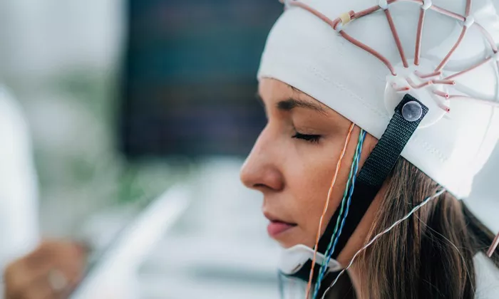
A new study led by researchers from the Institute of Science Tokyo suggests that electroencephalography (EEG) may offer a more accessible and effective alternative to functional magnetic resonance imaging (fMRI) for guiding transcranial direct current stimulation (tDCS) in the treatment of aphasia. The findings, published in NeuroImage on January 6, 2025, reveal an 80% agreement between EEG and fMRI in identifying brain regions activated during language tasks, and suggest that EEG-guided tDCS may improve treatment outcomes for individuals with language disorders.
Aphasia, a disorder that impairs language abilities, is commonly caused by damage to Broca’s area, a region in the brain responsible for speech production. While treatment options for aphasia remain limited, transcranial direct current stimulation (tDCS) has emerged as a potential therapeutic method. tDCS involves applying a low electrical current to the scalp to modulate neuronal activity, aiming to enhance or suppress brain functions.
Functional magnetic resonance imaging (fMRI) is currently the gold standard for mapping active brain regions, but its high costs and need for specialized equipment make it impractical for routine clinical use. This limitation has prompted scientists to explore alternative approaches, such as EEG, which is more affordable and accessible while offering a real-time measure of brain activity.
Professor Natsue Yoshimura and her team at the Institute of Science Tokyo sought to evaluate the potential of EEG as a tool for guiding tDCS in aphasia treatment. In their first experiment, they gathered both EEG and fMRI data from 21 healthy participants performing a picture-naming task. Participants were asked to quickly name objects shown in photographs, and the researchers compared the brain regions activated according to each method. An impressive 80% agreement was found between the EEG and fMRI data, confirming the validity of EEG as a tool for identifying brain regions involved in language processing.
In a follow-up experiment, the researchers tested the impact of EEG-guided tDCS on language performance by applying stimulation to brain regions identified by EEG, as opposed to the standard method that targets Broca’s area based on anatomical location. The results showed that EEG-guided tDCS significantly improved picture-naming speed compared to the traditional approach, suggesting that EEG may provide a more individualized and effective treatment.
Yoshimura’s team believes that their findings could pave the way for more personalized and accessible treatments for aphasia. “Our study provides the first indication that EEG-guided tDCS, accounting for individual differences in brain activity, has the potential to improve language function more effectively than conventional methods,” Yoshimura said. “These results also suggest that EEG-based analysis can identify brain areas relevant to specific cognitive tasks, further enhancing the rehabilitation process.”
As research in this area continues, EEG-guided tDCS may become a powerful tool in treating language disorders, offering a cost-effective, non-invasive alternative to traditional methods.
Related topic:
Persistent Poverty and Parental Mental Illness Linked to Increased Risk of Youth Violence
Study Reveals Genetic Links to Career Choices and Mental Health Traits
Obesity Drug Prescriptions Surge, Correlated with Online Interest

