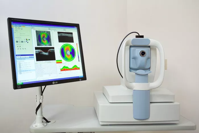
Optical Coherence Tomography (OCT) is a non-invasive imaging test that uses light waves to take cross-section pictures of your retina. The retina is the light-sensitive tissue lining the back of the eye. With OCT, your eye care professional can see each of the retina’s distinctive layers. This allows for the mapping and measurement of their thickness. These measurements help with diagnosis and provide treatment guidance for retinal diseases, such as age-related macular degeneration (AMD) and diabetic eye disease.
How Does OCT Work?
OCT is similar to ultrasound imaging, except that it uses light instead of sound. OCT can measure the thickness of the retina and the optic nerve fiber layer, which is important in glaucoma.
The technology provides detailed images of any structures within the eye that light can penetrate. The process is quick and painless, often completed in just a few minutes.
Benefits of OCT for Retinal Imaging
OCT has revolutionized the way eye care professionals diagnose and manage retinal conditions. It provides high-resolution images that are not possible with other imaging techniques.
This allows for early detection and treatment of eye diseases, potentially saving vision. OCT is also used to monitor the progression of diseases and the effectiveness of treatments.
Common Conditions Diagnosed with OCT
OCT is used to diagnose a variety of retinal conditions, including:
Age-Related Macular Degeneration (AMD): OCT can detect early signs of AMD, which is crucial for preventing vision loss.
Diabetic Retinopathy: This condition can cause swelling in the retina, which OCT can detect.
Glaucoma: OCT can measure the thickness of the optic nerve fiber layer, helping to diagnose and monitor glaucoma.
Macular Edema: Swelling in the macula, the part of the retina responsible for central vision, can be detected with OCT.
Retinal Detachments and Tears: OCT can provide detailed images of the retina, helping to identify detachments and tears.
Preparing for an OCT Scan
Before undergoing an OCT scan, your eye care professional will dilate your pupils with eye drops. This allows for a clearer view of the retina. The procedure is non-invasive and painless.
You will be asked to sit in front of the OCT machine and rest your chin on a support to keep your head steady. The machine will then scan your eye without touching it.
What to Expect During the OCT Scan
During the OCT scan, you will be asked to focus on a target while the machine captures images of your retina.
The process is quick, usually taking only a few minutes per eye. You may experience some sensitivity to light due to the dilation of your pupils, but this will subside after a few hours.
Interpreting OCT Results
The images produced by OCT are highly detailed and allow your eye care professional to see the different layers of the retina. This helps in identifying any abnormalities or changes in the retina.
Your doctor will explain the results and discuss any necessary treatments or follow-up appointments.
Advances in OCT Technology
OCT technology continues to advance, providing even more detailed images and new applications. Recent developments include:
Swept-Source OCT: This technology uses a longer wavelength of light, allowing for deeper penetration into the retina and choroid.
OCT Angiography: This non-invasive technique provides detailed images of the blood vessels in the retina, helping to diagnose conditions like AMD and diabetic retinopathy without the need for dye injections.
OCT in Research and Clinical Trials
OCT is not only a diagnostic tool but also plays a crucial role in research and clinical trials. It helps researchers understand the progression of retinal diseases and the effectiveness of new treatments.
OCT is used to monitor changes in the retina over time, providing valuable data for clinical studies.
Limitations of OCT
While OCT is a powerful tool, it does have some limitations. It may not be able to provide clear images if there is significant media opacity, such as cataracts or vitreous hemorrhage.
Additionally, OCT cannot replace a comprehensive eye exam and should be used in conjunction with other diagnostic tests.
Conclusion
OCT for retina is a groundbreaking technology that has transformed the diagnosis and management of retinal diseases. It provides detailed, high-resolution images that allow for early detection and treatment, potentially saving vision.
As technology advances, OCT will continue to play a crucial role in eye care, offering new possibilities for diagnosis, treatment, and research.
FAQs
What is OCT for retina?
OCT (Optical Coherence Tomography) is a non-invasive imaging test that uses light waves to take cross-section pictures of the retina, allowing for detailed visualization of its layers.
Is OCT for retina painful?
No, OCT is a painless and non-invasive procedure. It involves sitting in front of a machine that scans your eye without any physical contact.
How long does an OCT scan take?
An OCT scan typically takes only a few minutes per eye. The entire process, including preparation, usually takes about 15-30 minutes.
Can OCT detect all eye diseases?
While OCT is highly effective for diagnosing many retinal conditions, it may not detect all eye diseases. It is often used in conjunction with other diagnostic tests.
How often should I have an OCT scan?
The frequency of OCT scans depends on your eye health and any existing conditions. Your eye care professional will recommend how often you should have an OCT scan based on your individual needs.
Related topics:
3 Best Monofocal Lens for Cataract Surgery
Retina Transplant: A Comprehensive Guide to Restoring Vision

